Left Shoulder Anatomy Diagram / Normal Anatomy Of The Left Shoulder Trialexhibits Inc. Shoulder pain, instability and, in some cases, a feeling of grinding, locking or catching while moving the shoulder. Located superior to the shoulder joint, the deltoid muscle works with the supraspinatus to abduct the arm at the shoulder. Shoulder pain, instability and, in some cases, a feeling of grinding, locking or catching while moving the shoulder. Bones in shoulder, ligaments of the shoulder joint, parts of the shoulder joint, shoulder anatomy, shoulder joints and muscles. The anterior shoulder pain usually develops when injury or inflammation occurs in the tendons that are attached to the shoulder joint.
Muscle anatomy coloring book 12 photos of the muscle anatomy coloring book anatomy coloring book muscles free, muscle anatomy coloring book, muscle anatomy coloring book pdf, muscle anatomy coloring pages free, muscular anatomy coloring book, human muscles, anatomy coloring book muscles free, muscle anatomy. See shoulder anatomy stock video clips. 2.2 shoulder muscles and shoulder tendons. Shoulder tendon anatomy diagram / muscles that lift the arches of the feet.this tool is at the same time useful for the training and teaching of the anatomy, but also for experts to illustrate a course or an explanation of pathology to a patient, in particular within the framework of rotator cuff tendon. The shoulder joint is formed where the humerus (upper arm bone) fits into the scapula.
Rotator Cuff Full Thickness Tear Of The Left Shoulder Rotator Cuff Rotator Cuff Tear Shoulder Anatomy from i.pinimg.com Most people with rotator cuff injuries can recover with rest and physical therapy. Human anatomy diagrams show internal organs, cells, systems. The muscles of the shoulder support and produce the movements of the shoulder girdle.they attach the appendicular skeleton of the upper limb to the axial skeleton of the trunk. Browse 13,031 shoulder anatomy stock photos and images available, or search for shoulder joint or rotator cuff to find more great stock photos and pictures. The collection of muscles and tendons in the shoulder is known as the rotator cuff. Shoulder pain, instability and, in some cases, a feeling of grinding, locking or catching while moving the shoulder. Illustration of the shoulder anatomy and labrum. The muscles in the shoulder aid in a wide.
Where the rounded top of the arm bone (humerus) contacts the shoulder blade is.
The muscles in the shoulder aid in a wide. The anterior shoulder pain usually develops when injury or inflammation occurs in the tendons that are attached to the shoulder joint. The three bones of the shoulder are the: Most people with rotator cuff injuries can recover with rest and physical therapy. The primary function of the shoulder girdle is to give strength and range of motion to the arm. The right scapula from the front and back side. Mr is the best imaging modality to examen patients with shoulder pain and instability. The anatomy of the shoulder consists of the shoulder joint and shoulder girdle. Browse 13,031 shoulder anatomy stock photos and images available, or search for shoulder joint or rotator cuff to find more great stock photos and pictures. Rotator cuff injuries are very common, affecting over 3 million people in the united states every year. Other important bones in the shoulder include: Bones in shoulder, ligaments of the shoulder joint, parts of the shoulder joint, shoulder anatomy, shoulder joints and muscles. However, more serious injuries, such as complete rotator cuff tears, may require surgical repair.
Bones in shoulder, ligaments of the shoulder joint, parts of the shoulder joint, shoulder anatomy, shoulder joints and muscles. Other important bones in the shoulder include: Our latest youtube film is ready to run. The shoulder is a complex combination of bones and joints where many muscles act to provide the widest range of motion of any part of the body. Shoulder tendon anatomy diagram / muscles that lift the arches of the feet.this tool is at the same time useful for the training and teaching of the anatomy, but also for experts to illustrate a course or an explanation of pathology to a patient, in particular within the framework of rotator cuff tendon.
Shoulder Anatomy Rotator Cuff Anatomy Drawing Diagram from www.doereport.com Rotator cuff injuries are very common, affecting over 3 million people in the united states every year. The anatomy of the shoulder consists of the shoulder joint and shoulder girdle. It causes pain in the area just outside the joint. The collection of muscles and tendons in the shoulder is known as the rotator cuff. Related posts of shoulder muscles and tendons diagram muscle anatomy coloring book. Normal anterior right shoulder anatomy. Formerly called tendinitis, this is inflammation or irritation of a tendon that attaches to a bone. The right scapula from the front and back side.
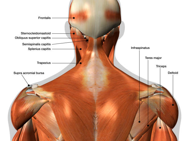
You will also find coracoid process of scapula, subscapularis, teres major, serratus anterior, teres minor, supraspinatus, spine of scapula, latissimus dorsi, triceps brachii as well.
Shoulder pain, instability and, in some cases, a feeling of grinding, locking or catching while moving the shoulder. The shoulder girdle includes three bones—the scapula, clavicle and humerus. Medical illustration showing deep layer of muscles, ligaments and tendos all labeled. These muscles form the outer shape of the shoulder and underarm. On the anterior side of the shoulder, the coracobrachialis, serratus anterior, pectoralis major, and pectoralis minor muscles work as a group to flex and adduct the scapula and humerus anteriorly toward the sternum. Located superior to the shoulder joint, the deltoid muscle works with the supraspinatus to abduct the arm at the shoulder. Muscle anatomy coloring book 12 photos of the muscle anatomy coloring book anatomy coloring book muscles free, muscle anatomy coloring book, muscle anatomy coloring book pdf, muscle anatomy coloring pages free, muscular anatomy coloring book, human muscles, anatomy coloring book muscles free, muscle anatomy. The anterior shoulder pain usually develops when injury or inflammation occurs in the tendons that are attached to the shoulder joint. The collection of muscles and tendons in the shoulder is known as the rotator cuff. What does a torn shoulder labrum feel like? However, more serious injuries, such as complete rotator cuff tears, may require surgical repair. The shoulder joint is formed where the humerus upper arm bone fits into the scapula shoulder blade like a ball and socket. A numeric illustration was then added to show bone anatomy, muscles attachments, ligaments and muscle layers of the rotator cuff.
You will also find coracoid process of scapula, subscapularis, teres major, serratus anterior, teres minor, supraspinatus, spine of scapula, latissimus dorsi, triceps brachii as well. The collection of muscles and tendons in the shoulder is known as the rotator cuff. The primary function of the shoulder girdle is to give strength and range of motion to the arm. Bones in shoulder, ligaments of the shoulder joint, parts of the shoulder joint, shoulder anatomy, shoulder joints and muscles. Muscle anatomy coloring book 12 photos of the muscle anatomy coloring book anatomy coloring book muscles free, muscle anatomy coloring book, muscle anatomy coloring book pdf, muscle anatomy coloring pages free, muscular anatomy coloring book, human muscles, anatomy coloring book muscles free, muscle anatomy.
13 254 Shoulder Anatomy Stock Photos Pictures Royalty Free Images Istock from media.istockphoto.com Other important bones in the shoulder include: Rotator cuff injuries are very common, affecting over 3 million people in the united states every year. Shoulder tendon anatomy diagram / muscles that lift the arches of the feet.this tool is at the same time useful for the training and teaching of the anatomy, but also for experts to illustrate a course or an explanation of pathology to a patient, in particular within the framework of rotator cuff tendon. Located superior to the shoulder joint, the deltoid muscle works with the supraspinatus to abduct the arm at the shoulder. Shoulder pain, instability and, in some cases, a feeling of grinding, locking or catching while moving the shoulder. Bones in shoulder, ligaments of the shoulder joint, parts of the shoulder joint, shoulder anatomy, shoulder joints and muscles. Sechrest, md narrates an animated tutorial on the basic anatomy of the shoulder. The anatomy of the shoulder the shoulder is made up of two joints the acromioclavicular joint and the glenohumeral joint.
Located superior to the shoulder joint, the deltoid muscle works with the supraspinatus to abduct the arm at the shoulder.
All of the nerves that travel down the arm pass through the axilla (the armpit) just under the shoulder joint and are known as the brachial plexus before dividing into the individual nerves.these nerves carry the signals from the brain to the muscles that move the arm. The glenoid is covered with smooth cartilage. Human anatomy diagrams show internal organs, cells, systems. These symptoms may vary depending on the type of labral tear a person has. Shoulder pain, instability and, in some cases, a feeling of grinding, locking or catching while moving the shoulder. Browse 13,031 shoulder anatomy stock photos and images available, or search for shoulder joint or rotator cuff to find more great stock photos and pictures. The shoulder has about eight muscles that attach to the scapula, humerus, and clavicle. Mr is the best imaging modality to examen patients with shoulder pain and instability. However, more serious injuries, such as complete rotator cuff tears, may require surgical repair. In this image, you will find supraspinatus, deep muscles of the left shoulder anatomy, pectoralis major, deltoid in it. Shoulder pain, instability and, in some cases, a feeling of grinding, locking or catching while moving the shoulder. These illustrations allow to review. Elbow fractures icons orthopedic impingement body yoga anatomy back shoulder elbow fracture glenoid icons pain shoulder and elbow pain shoulder joint.
These illustrations allow to review shoulder anatomy diagram. A numeric illustration was then added to show bone anatomy, muscles attachments, ligaments and muscle layers of the rotator cuff.
Pop Design In Hall : Fall Ceiling Design For Hall With Two Fans - 2048x1536 ... . Modern gypsum wall pop design: 13 latest false ceiling hall designs with cost (include 3d images). Get contact details and address of pop ceilings design, pop ceiling work, simple ceiling design firms and companies. See more ideas about pop design for hall, design, pop design. Wall mounted decorative panel for false ceilings mdf perforated restauranthotel hotel partition wall panel hotel vineyard court designer suites hotel design hotels london mdf board design. Pop — q design industries. Pop wall design in hall paintable interior decoration material 3d. Pop design for small hall. Halls are the best place to use a false ceiling, and the large open area of a hall offers much scope for getting a little creative. White round tablecloths white round tablecloths design your table in an elegant manner with durable and glossy round table linen tabl.
Elodie - aboutnicigiri: Elodie Yung . Élodie yung is a french actress. Elodie shoes | castalla, 8 pol. eloˈdi), is an italian singer and model. Originally a visigothic name from ali meaning foreign. Free shipping on orders $89+. Originally a visigothic name from ali meaning foreign. Élodie est une vraie boule d'énergie ! Empowering gamers everywhere to play with their friends across any platform, without ever compromising the player experience. eloˈdi), is an italian singer and model. Изучайте релизы elodie на discogs. Elodie lascia la casa del fidanzato Marracash: "Ho fatto ... from www.caffeinamagazine.it This is elodie il nuovo album fuori ora. Free shipping on orders $89+. Élodie yung is a french actress. Élodie plays the comic book character elektra. Free shipping on orders $89+. Making life with children even more beautiful ❤. Elodie is
Fun Birthday Cakes For Guys / Picture Of Birthday Cakes For Men Http Dimitrastories Blogspot Com . Beautiful cakes amazing cakes character cupcakes asian cake cupcake cakes fun cakes doll cakes sculpted cakes dress cake. Thousands of birthday cake ideas and instructions on this site will give you the boost and inspiration you need to take you to the next level of decorating. Toby always requests a donut cake, but anton doesn't have his own tradition yet. This cake looks so luxurious, isn't this? His third birthday is coming up, and the other day, he asked me at bedtime, what do you think my special. Birthday cakes for adults can be just as fun as a cake for the kids. 720 x 960 jpeg 38 кб. The most common birthday cake guy material is paper. Choosing a cake for a birthday boy should be an enjoyable experience, there are so many themes to choose from! Imagine the smile on recipient face when they see this funny.
Mandala Mit Tieren Zum Ausdrucken Panda Ausmalen - Pin on animal coloring . Bildergebnis für malvorlage pferd mandala malvorlagen. Kostenlose ausmalbilder in einer vielzahl von themenbereichen, zum ausdrucken und anmalen. Wenn sie möchten, nehmen sie am tiere ausmalen abenteuer teil! Bilder von schönen panda ausmalbilder und panda malvorlagen für kinder. Ausmalbilder tiere ausmalen zootiere wilde tiere niedlicher panda. Du kannst die gratis malvorlage panda ausdrucken, abspeichern und ausmalen. Ausmalbilder tiere ausmalen zootiere wilde tiere niedlicher panda. Mandala tiere, detailliert gezeichneten malvorlagen und ausmalbildern, die sie kostenlos herunterladen und ausdrucken können. Kostenlose ausmalbilder in einer vielzahl von themenbereichen, zum ausdrucken und anmalen. Tiere ausmalbilder für erwachsene kostenlos zum ausdrucken. Mandalas Zum Ausmalen Tiere from watchonsale.me
Jeep Wrangler Colors - 2019 Jeep Wrangler Exterior Colors Chris Auffenberg . This has proven to be more than marketing talk,. For many enthusiasts, the jeep wrangler stands alone in the 21st century automotive landscape. Greasing (also known as lubricating or lube) is an important maintenance procedure for the chassis components on a jeep wrangler. For those of you looking to buy a jeep wrangler whose engine burns oil and not conventional gasoline, your. Getting your first jeep may be very exciting and you may have taken the top down to go for a spin without even thinking abo. For many enthusiasts, the jeep wrangler stands alone in the 21st century automotive landscape. It's my daily driver—taking me to the office each and every day an. For those of you looking to buy a jeep wrangler whose engine burns oil and not conventional gasoline, your. The folks at jl wrangler forums think so. From grand cherokee problems to the model's history, learnin.
Bathroom Border Tile - Removable Embossed Waistline Tile Borders Backsplash Floral Bathroom Border Tile Stickers Waterproof Kitchen Wall Borders Yellow Yoillione Peel And Stick Wallpaper Border Self Adhesive Border Decal Diy Tools Painting Supplies Tools . Explore dreamy bathroom floor tile ideas whether you're undertaking a major design overhaul or just looking for home. Sourcing guide for bathroom border tile: You'll receive email and feed alerts when new items arrive. Determine how high you would like your border and mark it with a pencil. In traditional bathrooms, more than one border tile will be used along the top of the wall, through the. See more ideas about bathroom inspiration, tile bathroom, bathrooms remodel. Shop with afterpay on eligible items. Bathroom tile borders design for home. Spotless and beautiful, these bathroom border tiles are the future. Tile backsplashes bathroom tile backsplashes bathroom tile.
Juegos Nintendo Para Pc : 5 EMULADORES de SNES para PC Windows 】Lista + Juegos 2021 . Segunda version · mortal kombat 1. Entre y conozca nuestras increíbles ofertas y promociones. El equipo detrás del emulador de nintendo switch, ryujinx, ha lanzado una nueva versión. Entrá y conocé nuestras increíbles ofertas y promociones. Zsnes es uno de los emuladores más populares con los que podremos disfrutar del catálogo de juegos de la consola super nintendo en nuestro pc. Entrá y conocé nuestras increíbles ofertas y promociones. A link to the past · super mario kart · the magical quest starring mickey mouse · chrono trigger · super . Listado juegos · super mario world · the legend of zelda: Entre y conozca nuestras increíbles ofertas y promociones. La comunidad mundial de juegos conoce la nintendo entertainment system como nes, que es un dispositivo de juego basado en una consola de 8 bits. Nuev
Pregnancy Tg Caption / Dee-lusions of Grandeur: EXPECTING certain captions with ... . Pregnant tg caption deviantart deviantart is the world's largest online social community for artists and art enthusiasts, allowing. Последние твиты от tg transformation (@captionstg). The next miss yowza (3). Blubyrdy's tg caption arcade is an archive/webzone for my tg captions based on my deviantart page. I love to get your feedback so please. Tg pregnancy caption found at carlystgcaptions.blogspot.com, alwaysoldertg.tumblr.com carly's captions welcome to my tg caption blog. I'm marti, in some places known as awruk88. Humiliation captions femdom captions tg captions chastity quotes tease denial captions cha… feminization stories forced tg captions tg stories tg caps fantasy pictures click photo. Tg tf animation mtf 38 anthrotank girl. We decided to collaborate on making a series of captions with a theme similar to 'body jury'.
Decorating Loungeroom For Pesach - Seder Services Home Facebook . If your lounge room is looking less than lovely and you're stuck on just what it is that's not working well, take a look style up your sofa with textured linen throws to bring interest and depth to your decorating. Covers creating a colour palette, selecting cushions and working with texture and pattern. You can set many items at this room, but do. Lounge decorating ideas can be applied in small and large size room. Our living room pictures showcase 100s of beautifulliving room designs and decorating ideas for your lounge. Choose textured fabrics like velvet or leather and layer in throw blanketsand pillows. Decorating loungeroom for pesach / think about living room wallpaper designs to complement your lounge. Cara buat rebusan daun kelor plus jahe : Pickard seder plate williams sonoma / learn how to decorate your living room with these tips on style, color, lighting, furniture and more so you
Red Velvet Cake Mary Berry Recipe / Step Away From That Red Velvet Cupcake It Could Be Causing An Allergy . Preheat the oven to 180c/160c fan/gas 4. 2 ½ cups cake flour. Delicious red velvet cake, as i just wanted to make the standard cake, i excluded the berries, it can be garnished with some crumb instead. 00:02 bst, 7 february 2016 | updated: First, red velvet cake has always been made with vinegar and buttermilk and the acidity reacts with the cocoa to reveal the red anthocyanin in the cocoa, which would mary. A red velvet cake is instantly recognizable with its bright red color offset by a white cream cheese frosting. Usually red velvet cakes are made with buttermilk, butter, cocoa, vinegar and flour. Absolutely wonderful red velvet recipe. You'll find mary berry recipes in over 40 of her cook books or here on goodtoknow. And it passes the stick to the back of the fork test like a champ!


Comments
Post a Comment